
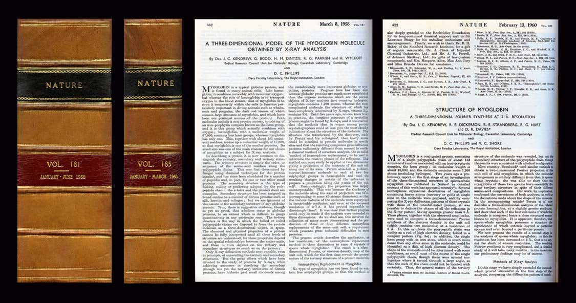
A Three-Dimensional Model of the Myoglobin Molecule Obtained by X-ray Analysis in Nature 181, 1958, pp. 662-666 WITH Structure of Myoglobin: A Three-Dimensional Fourier synthesis at 2 Å Resolution in Nature 185, 1960, pp. 422-27
1st Edition. Two volume first edition of a milestone in the history of structural biology, the first protein structure ever discovered. Kendrew’s papers present the first solution of the three-dimensional molecular structure of a protein. “Kendrew’s discovery was one of the greatest landmarks in the history of molecular biology” (Jeremy Norman History of Science). Along with his colleague Max Perutz, Kendrew received the 1952 Nobel Prize for this work.
Two discoveries laid the foundation for Kendrew’s work: “first, that the positions of atoms in a crystallized substance could be determined from X-rays passing through, and scattered by, the crystal; second, that the approach could be applied to very simple biological molecules (Garwin, Century of Nature, 88).
The goal of Kendrew’s team, composed mostly of physicists, was to understand how a biological molecule such as a protein works, beginning with how it is built. The papers they produced (both offered here) are “the outcome of a truly Herculean task. When the project began, there were no automated instruments or digital computers for generating or analyzing the huge amounts of data necessary. Every step had to be carried out by hand… many repeated thousands of times” (ibid, 89).
Kendrew selected myoblobin as a protein because it abundant in the tissue in which it operated, is an oxygen-binding protein from muscle, and is very stable. His next step “was to measure the amplitudes of the ‘reflections’ – the scattered X-ray beams – produced when the crystals were exposed to X-radiation. Following this, he add up the measured reflections, in a process called Fourier summation, to produce an image of the molecules in the crystal lattice. “Photographic film was used to record the reflection amplitudes, thousands of them, and then teams… were trained to estimate their values by comparison with a known scale… The process took years.
“The final step was the Fourier summation itself. Kendrew… [carried] out an earlier trial at lower resolution [and did] the summation by hand… For the more than 10,000 reflections that had to be added up at 2 angstrom resolution, hand summation was impossible… Technology came to the rescue: the first high-resolution protein structure ever determined was calculated on the first electronic computer ever built in Britain.
“The resulting electron-density map looked impossible to interpret, [but] an ingenious system was devised for building the atomic model of the protein: the map was produced as a set of contoured topographic plots on sections through the crystal lattice; a three-dimensional grid was overlaid onto the map so that atomic coordinates could be measured with respect to three axes, x, y, and z, at right angles to each other; and wire rods were constructed whose height was proportional to the z-coordinate of each feature of density. Brass models were fabricated for each amino acid in the protein, and these were fitted by hand into the ‘forest’ of wires and secured by clamps” (ibid 90).
The structure that resulted was not one anyone was prepared for – “the seeming irregularity of the overall polymer fold and the remarkable fact that the heme iron atom where oxygen bound was buried completely inside the protein” – and yet there were striking patterns, including the polymer chain folded into a series of eight segments (ibid). “After almost one hundred years of speculation, one could now see what a protein looked like in atomic detail” (ibid). Item #907
CONDITION & DETAILS: 4to. (10.25 x 7.5 inches; 256 x 188mm). Ex-libris: bearing only a small circular stamp on the rear of the title page. Handsomely rebound in half calf over the original cloth covered boards. 5 raised bands at the spine, each gilt-ruled. Red and black morocco spine label, gilt-lettered. Tightly and very solidly bound. Bright and very clean throughout. Very good to near fine condition.
Price: $4,000.00